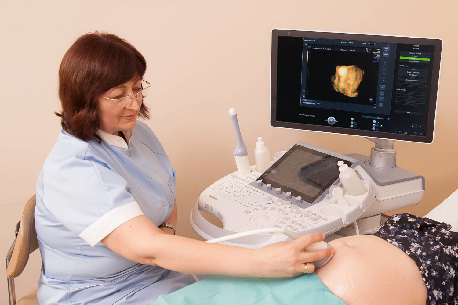
Foetal ultrasound includes the assessment of the structure of the lesser pelvis, determining pregnancy, time of childbirth, development of the foetus, position of the placenta and umbilical cord, which is done by using high class ultrasound devices, as well as observation of the spatial image of the foetus and superficial assessment of organs in real time, by using 2, 3 or 4 dimension (4D) ultrasound with colour Doppler. The examination is available at any gynaecological consultation.
2D ultrasound examination is an indispensable part of basic examination. The principal image of the examination is created by ultrasound, as it passes tissue of different density, depth, blood supply and localisation. However, the possibilities of modern technology have enabled to direct the ultrasound into most different directions and, as the computer processes the obtained data, a spatial image is received, which is currently known as 3D ultrasonography. As technologies are upgraded, spatial image in real time can be obtained, which is called 4D ultrasonography.
National Health Service (NHS), insurance company and paid services are available.