Description

The Author: Veselības centrs 4

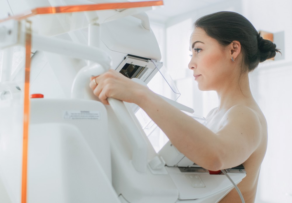
Breast cancer is the most common oncological disease in the world, which claims lives of many women. However, prompt diagnosis and detection of the diseases at early stages enables successful treatment thereof. The most precise diagnostics of breast health nowadays can be performed by using digital mammography devices with tomosynthesis.  The newest devices enable not only faster, more comfortable, but also deeper examination of breasts by means of mammography.

Two examinations are performed during one – digital mammography and an in-depth examination, which is ensured by tomosynthesis. This examination method is considerably more precise and enables earlier detection of changes and possible pathology, because specialists are given an opportunity to view the image layer by layer, to visualise structure in each layer separately and to view a moving image from 3 sides. This examination also reduces the need for a repeated examination. The device recognises and automatically labels suspicious structures by giving special warnings and pointing physician''s attention to them.

The technology of tomosynthesis is considered the greatest achievement in the development of mammography method over the last 40 years. Tomosynthesis ensures special early diagnosis advantages for patients with dense breast structure, which most commonly occurs in young women. The ability of earlier mammography techniques in such cases were rather limited and women had to make other examinations - ultrasound or magnetic resonance examinations. However, to specify the diagnosis, physicians more and more frequently tend to recommend ultrasound examination as well.

Patient benefits:

  • Higher comfort during examination. The examination is completely painless for a woman, because it is performed with a mobile paddle that adjusts to the shape of the breast. To ensure that the examination is performed successfully, the device itself registers the force that needs to be applied to the paddles of the device in accordance with the physiological characteristics of the patient''s body, as well as the examination field of the breast is determined automatically by adjusting it closer tot he chest. He entire gland up to the nipple is included.
  • The radiation dose is reduced thanks to the short duration of examination.
  • The result of examination is stored in the electronic patient data base, which enables comparison of dynamic change between every new examination and the previous one.

During this 3D examination, very small parts and regions of breast, which do not overlap, can be visualised and assessed. Thus, a considerably higher precision of diagnostics can be obtained faster and safer, which could not be possible to set up in a 2D image. This examination also reduces the need for a repeated examination. The device recognises and automatically labels suspicious structures by giving special warnings and pointing physician''s attention to them.

Tomosynthesis – an imaging technology as a result of which the examination of breast is performed in three-dimensional images, where the images are created from different projection angles. The projection of the image is reconstructed by reproducing the cross-section of the breast with the interval of 1 mm.

Patients with National Health Service (NHS) invitation letters, as well as patients with the referral of a general practitioner can undergo digital mammography at Veselības centrs 4 Ltd., by paying only the co-payment determined by the state. Performance of digital mammography with tomosynthesis will be a special benefit, because examination of two breasts are performed within the framework of standard mammography!

National Health Service (NHS), insurance company and paid services are available.

The service is available in the following branches

Sign up