Magnetic resonance imaging (MRI) is a visual examination method that is completely harmless for human health. Electromagnetic field and radio frequency wave impulses are used to obtain the magnetic resonance image.
One of the most precise diagnostic examination methods, which allows obtaining cross-section images of human body and organs. This method is characterised by high precision and in many cases provides more comprehensive information than computed tomography.
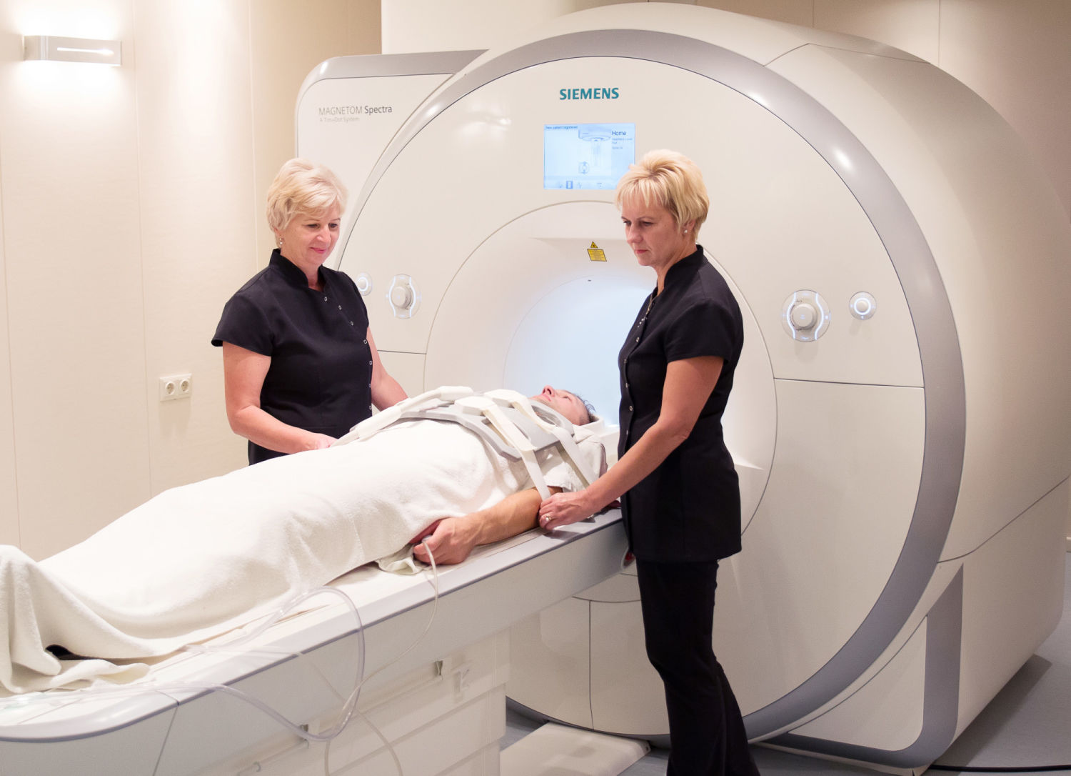
At the MRI office of Orto Clinic of Veselības centrs 4, Ltd. a Siemens MAGNETOM Spectra MR (3T) device is installed, which enables the highest quality and speed of examinations, which is especially important in the event of serious trauma and diseases, when patients find it difficult to remain motionless for a long period of time.
Siemens MAGNETOM Spectra MR is special in terms of its capacity to perform simultaneous entire body scan. The possibility of this examination provides especial benefit for oncology patients, since detection of early metastases is impossible without entire body scan.
It is also indispensable for diagnostics of small joints and organs, as well as in the event of multiple sclerosis as it enables visualisation of several foci, which cannot be done by other standard MRI devices.
Magnetic resonance imaging is used for examination of:
- Brain;
- Soft tissue of the head and neck;
- Blood vessels;
- Vertebral column and spinal cord;
- Bones and joints;
- Muscle and other soft tissue.
National Health Service (NHS), insurance company and paid services are available.